How to treat wound malodour?
Identify, manage and neutraliseTo treat wound malodour effectively, it is important for clinicians to identify and manage the underlying cause first. Any wound can become malodorous, especially if infection, devitalised tissue or high levels of exudate are present. [1] In some cases, multiple factors may be causing the malodour and clinicians must treat them simultaneously to ensure the best patient outcomes.
Although there is limited guidance on managing malodour, using a pathway that prioritises the identification and treatment of the cause[s] of the odour can be a useful part of a holistic and individualised care plan. Treatment should also focus on neutralising the malodour to minimise the physical and emotional distress of patients and their carers.
How is wound infection treated?
Infection and bacteria are one of the main causes of wound malodour. [2] Bacteria that normally live harmlessly on the skin and in the patient’s environment are drawn to wounds, where the nutrient-rich wound bed and exudate create an ideal environment for bacterial growth. The immune system usually responds to control this growth, but if the bacteria are extremely virulent or the immune response is insufficient, infection can occur. [3] To treat wound malodour caused by infection, follow local policy and consider the infection continuum (Table 1), developed by the International Wound Infection Institution (IWII).
Table 1: Treatment of infection using the wound infection continuum
| Position on wound continuum | Signs and symptoms | Signs and symptoms |
|---|---|---|
| Local infection | Overgranulation, bleeding friable tissue, increased exudate, delayed healing, wound breakdown, erythema, warmth, swelling, increased pain and malodour | Cleansing and debridement. Consider treatment with topical antimicrobial, i.e., silver, iodine, PHMB, honey |
| Spreading infection | All of the above plus: spreading erythema into surrounding tissues >2cm crepitus, wound breakdown and swelling of the lymph glands | Cleansing and debridement. Commence systemic antibiotics and consider adjunctive treatment with topical antimicrobial agent. Swab wound using Levine technique to correctly identify organism present and antimicrobial sensitivities |
| Systemic infection | All of the above plus: malaise, lethargy, loss of appetite, fever/pyrexia, septic shock, organ failure, death | Urgent medical referral needed and start of systemic antibiotics. Topical antimicrobials may be used as adjunctive therapy |
How is devitalised tissue treated?
The terms devitalised and necrotic are often used interchangeably to describe dead tissue on the wound bed. Tissue can die when blood supply to the area is lost, blocked arteries, trauma, infection or prolonged pressure. Devitalised tissue can delay wound healing because it acts as a source of nutrition for bacteria, which increases the risk of infection. [4]
Debridement is the act of removing devitalised tissue. There are several types of debridement, including autolytic, mechanical, biological, enzymatic, surgical and sharp debridement. A holistic assessment of the patient must be performed before debridement to determine:
- Wound healing potential post-debridement
- Appropriate type of debridement
- Patient preferences and priorities. [5]
It is important to recognise that debridement may not always be appropriate or suitable for certain wounds. Situations where debridement may not be appropriate include end-of-life patients or those with advanced necrosis due to arterial disease.

Autolytic debridement
Autolytic debridement is when the body’s enzymes in wound exudate break down devitalised tissue. This type of debridement is often slow, but can be accelerated via other clinical interventions, including the use of dressings.
Mechanical debridement
Mechanical debridement uses force to remove devitalised tissue. Traditionally, clinicians use a ‘wet-to-dry’ method for this procedure. They place a wet gauze on the wound and let it dry so it adheres to the top layer of the wound bed. As they remove the gauze, the devitalised tissue is pulled away. Although this method is no longer recommended because of pain and additional wound trauma, there are newer techniques, including using moistened gauze or monofilament fibre pads to clean away devitalised tissue. [6] [7]
Biological debridement
This method is also known as larval debridement therapy (LDT). The procedure involves placing larvae directly on the wound, where they feed on devitalised tissue and bacteria. The length of treatment depends on the wound type and condition.
LDT can be controversial for several reasons, including misconceptions about the treatment, as well as ethical and aesthetic objections. [8]
Surgical and sharp debridement
These methods are performed using sterile curettes, scalpels, forceps or scissors.
Surgical debridement is usually reserved for larger wounds and involves the removal of all devitalised tissue to create a healthy, bleeding wound. On the other hand, clinicians usually carry out sharp debridement when the devitalised tissue starts to separate from the healthy tissue.
It is recommended that only trained clinicians perform surgical and sharp debridement. [9] These methods are not advisable for malignant fungating wounds (MFWs) due to the risk of bleeding and cell seeding. [10]
How to manage malignant fungating wounds?
Malignant fungating wounds (MFWs) are a distressing cancer-related complication. They occur when a cancerous tumour close to the surface of the skin breaks through and creates an open wound or originate directly from primary cancers of the skin (ie., squamous cell carcinomas).
When cancer is the cause of a wound, clinical interventions from specialist oncology, dermatology and plastic surgeon consultants are required to manage or remove the cancerous cells to promote healing. When curative treatment is not possible, a palliative approach that focuses on minimising patient distress caused by wound malodour is recommended. [11]

How to manage generalised wound odour and exudate?
Wound malodour is often accompanied by moderate-to-high levels of exudate. Clinicians should first identify and treat the cause[s] of the excessive exudate. This may include the use of compression or referral for additional clinical services (i.e., lymphoedema).
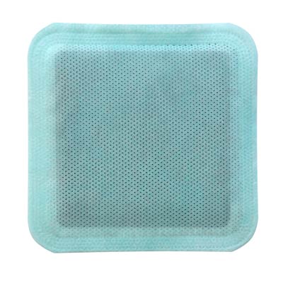
Dressing selection should consider exudate levels, malodour and patient comfort. Where the malodour is caused by heavy exudate, a multipurpose dressing that can absorb and retain fluid, while helping to reduce odour.
References:
- Pramod, S (2025) Impact of Wound Malodour on Patients: How to Assess and Manage. J Community Nurs 39(1): 18-25
- Ibid
- International Wound Infection Institute (2022) Wound Infection in Clinical Practice. Wounds International, London
- Mayer O, Dieter, et al (2024) Best Practice for Wound Debridement. Journal of Wound Care 33 (6b): 1-32
- Vowden, K, Vowden P (2011) Debridement: Made Easy. Wounds UK 7(4): 1-4
- Fleck, C A (2009) Why “wet to dry”? Journal of the American College of Certified Wound Specialists. 6;1(4):109-13
- Mayer O, Dieter, et al (2024) Best Practice for Wound Debridement. Journal of Wound Care 33 (6b): 1-32
- Atkin, L, Acton, C, Edmonds, M, et al (2020) The Role of Larval Debridement Therapy in the Management of Lower Limb Wounds. Wounds UK. London
- Mayer O, Dieter, et al (2024) Best Practice for Wound Debridement. Journal of Wound Care 33 (6b): 1-32
- Ibid
- Ousey K, Pramod S, Clark T et al (2024) Malignant wounds: Management in practice. London: Wounds UK.
- Pramod, S (2025) Impact of Wound Malodour on Patients: How to Assess and Manage. J Community Nurs 39(1): 18-25
Disclaimer:
Please note that while every effort is made to ensure the accuracy of the content presented, it is purely for educational purposes only and is not a substitute for professional medical advice.
