What are the 10 main precautions for
safer suction catheter use?
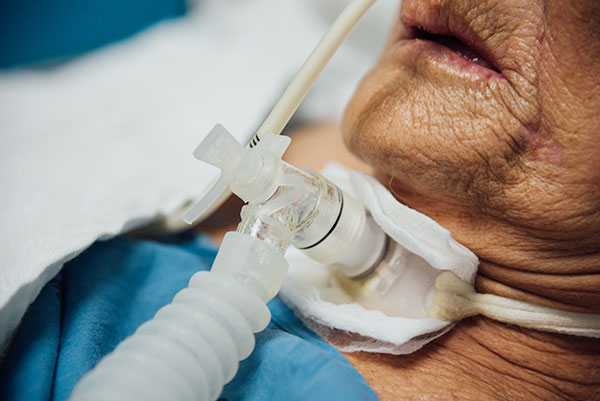
Suctioning is a procedure that is carried out to aspirate secretions from the trachea to prevent a potentially life-threatening blockage in the airways.
Although suctioning is a lifesaving procedure, it can lead to a range of complications, including hypoxia and an irregular heart rate.
What can we do to ensure safer suction catheter use?
Due to the possibility of complications, it is important for healthcare professionals to perform suctioning carefully and appropriately. There are a variety of measures that can be taken to ensure and protect the safety of both patients and healthcare professionals alike.
Carefully assess the patient
Suctioning is an invasive procedure with a range of potential adverse effects. It should be performed infrequently and following a thorough assessment that determines the clinical indications and needs of the patient. [1]
The assessment should examine different aspects of the respiratory system.

Audible symptoms:
• Wheezing
• Whistling
• Coughing
• Shallow breaths
• Cackling
Visible symptoms:
• Visible secretions in the trachea
• Cyanosis
Physical symptoms:
• Decrease in oxygen saturation
• Increased airway pressure
• Tachycardia
• Irregular heartbeat
• Restlessness
After the assessment has been completed, the clinician should evaluate all the symptoms in order to ascertain the need for patient suctioning.
Communicate with the patient
Once suctioning is advised, it is important to explain the procedure to the patient. Communicating with the patient is critical because:
• Allows the clinician to obtain consent from the patient.
• Helps to alleviate the anxiety of the patient and fosters cooperation, facilitating the removal of secretions.
• Enables the clinician to monitor the patient’s wellbeing before suctioning, between passes, and after the procedure.
Use PPE
An assessment of the risk of exposure to infectious agents and bodily fluids should be performed before each procedure. Personal protective equipment (PPE) that is suitable for that procedure should be worn to minimise the risk of contamination and infection. [2] Examples of PPE include:
• Gloves
• Aprons
Using PPE will ensure safer suction catheter use for both the patient and the clinician.
Use the correct suction catheter size
It is recommended that suction catheters not exceed 50% of the internal diameter (lumen) of the tracheal tube. [4] This minimises the risk of mechanical trauma, which can cause injury and bleeding that can lead to infection. An appropriately sized suction catheter may also prevent large amounts of oxygen being evacuated from the lungs all at once, causing a significant drop in oxygen levels. [5]
Suction catheters that are too small in diameter also carry their own set of complications. If the suction catheter is small, it will be inadequate at removing secretions effectively. This increases the risk of trauma as more passes will need to be performed to remove the secretions. [6]
Preoxygenate patient
The oxygen levels of the body can be adversely affected by suctioning. These fluctuations may result in hypoxia, hypoxemia and even atelectasis (partial or full collapse of the lungs). [7]
Administering oxygen before and between passes is advised to mitigate the effects of oxygen aspiration during suctioning. Pre- and post-oxygenation should be carried out using a ventilator and last a minimum of 30 seconds. It is important to check the patient’s oxygen saturation throughout the procedure. [8]
Limit the frequency of suctioning
Suctioning is a potentially dangerous procedure if performed too frequently and poorly. Each pass of the suction catheter can have a negative effect on a patient’s intracranial pressure, oxygen saturation, heart rate and blood pressure. [9]
The more a patient is suctioned, the higher the chances of adverse effects taking place. The patient’s clinical condition should dictate the interval between individual suctioning procedures.
The assessment will determine the frequency of suctioning based on the amount of secretions, the patency of the airways and the clinical status of the patient.
Duration of suction catheter passes
It is recommended that suction time be limited to 15 seconds per pass. [10] Time should be left between passes to aid respiratory recovery.
It is generally advised that no more than 3 passes should be carried out in one session. [11]
Appropriate suction pressure
Selecting a suction catheter with multiple holes, known as ‘eyes,’ at the tip is one of the keys to controlling the pressure. Studies show that suction catheters with two holes are better at distributing pressure and reducing trauma during the procedure. [12]
Another important consideration for safer suction catheter use is the amount of negative pressure employed. Clinicians must strike a balance between removing the secretions and limiting trauma to the airways. For this reason, suction pressure should be set as low as is reasonably possible to remove the secretions.
The amount of pressure required by a patient depends on a range of factors, including:
- The secretions
- Age
- Size
- Clinical condition
It is, however, generally recommended that suction pressure not exceed 200 mmHg for adults. [13]
Monitor the depth of suction
Another key factor to consider for safer suction catheter use is the depth of insertion. The suction catheter should be inserted into the trachea, no further than the carina. Signs that the appropriate depth of insertion has been reached include feeling resistance against the suction catheter and light coughing. [14]
Pushing the catheter beyond the carina can lead to a series of complications, including mucosal trauma and arrhythmias. [15]
Document
It is critical to document the findings of any clinical assessment and to report on patient progress. This will not only improve communication among clinical staff, but also ensure consistency in treatment. Examples of information that should be documented include:
- Size of endotracheal tube
- Patient condition
- Volume of secretions
- Colour and odour of secretions
- Size of suction catheter used
- Suction pressure used
- Number of passes
- Patient responsiveness to suctioning
- Complications of suctioning
References:
- Leeds Teaching Hospitals NHS Trust. Tracheal Suctioning – Clinical Protocols for Adults 2022. http://www.lhp.leedsth.nhs.uk/detail.aspx?id=3152 [accessed 30th September 2023]
- NHS England. National Infection Prevention and Control Manual for England. https://www.england.nhs.uk/national-infection-prevention-and-control-manual-nipcm-for-england/ [accessed 20th September 2023]
- Leeds Teaching Hospitals NHS Trust. Tracheal Suctioning – Clinical Protocols for Adults 2022. http://www.lhp.leedsth.nhs.uk/detail.aspx?id=3152 [accessed 30th September 2023]
- Ibid
- Ibid
- Ibid
- Ibid
- Ibid
- University Hospitals Sussex NHS Foundation Trust. Guideline for Adult Endotracheal Suction in Critical Care. https://www.bsuh.nhs.uk/library/wp-content/uploads/sites/8/2022/08/Endotracheal-suction-2022.pdf [accessed 30th September 2023]
- Ibid
- Ibid
- Ibid
- Ibid
- http://www.lhp.leedsth.nhs.uk/detail.aspx?id=3152 [accessed 30th September 2023]
- Ibid
- Ibid
- St George’s Healthcare NHS Trust. Adult, Paediatric and Neonatal Suction Policy. https://www.stgeorges.nhs.uk/wp-content/uploads/2013/11/Clin_4_07Suction.pdf [accessed 29th September 2023]
Resources:
Product website:
Other related articles:
Disclaimer:
Please note that while every effort is made to ensure the accuracy of the content presented, it is purely for educational purposes only and is not a substitute for professional medical advice.
